Mechanism Of Tilmicosin's Antiviral Activities In Cases Of PRRSV
Ulrich KleinPorcine respiratory reproductive syndrome virus (PRRSV) is the aetiological agent of one of the most persistent viral diseases affecting pigs globally.
Novel PRRSV variants are present worldwide which are highly pathogenic and aggressive, causing severe respiratory symptoms, and high mortality and abortion rates. Commercially available vaccines provide insufficient immune response to cover the full breadth and diversity of existing PRRSV genotype variants.
Tilmicosin accumulates in pulmonary alveolar macrophages (PAMSs) to high concentrations1.
For effective prevention and control of current and future PRRSV variants, the use of tilmicosin is important based on its anti-PRRSV activity.
In a new study, the mechanism of the anti-PRRSV effect of tilmicosin was investigated in vitro (measuring pulmonary alveolar macrophages (PAMs) and monkey kidney Marc-145 cells) and in vivo (in a piglet challenge study)2.
Role of macrophages and cellular factors in PRRSV infections
Macrophages are the key target cells for PRRSV infections and the main site of PRRSV replication. Several entry mediators and receptors (CD169; MYH9; CD209; CD163) for PRRSV have been identified in macrophages. The key cellular receptor for PRRSV infection is CD163.
CD163 is the main cellular receptor necessary for PRRSV entry and is sufficient to create PRRSV infections3.
Studies into PRRSV antiviral activity
In the in vitro study2, the effect of tilmicosin against currently prevalent PRRSV strains (N34, N30, HP PRRSV-2 and PRRSV-1) in PAMs and MA104-derived monkey kidney Marc-145 cells were determined by using increased tilmicosin concentrations (50-150 µg/ml).
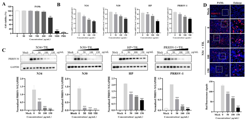
Figure 1. Inhibition of currently prevalent PRRSV infection by tilmicosin in PAMs in different tilmicosin concentrations (in vitro study results). Statistical significances are denoted by * p<0.05; ** p<0.01; *** p<0.001
As shown in Figure 1B and determined by real-time RT-PCR testing, tilmicosin (50-150 µg/ml) reduces (p<0.05) the different PRRS virus types present in the PAMs (level of PRRS virus mRNA determined) dose-dependently. Western-blotting analysis results (Figure 1C) indicate that N protein expression in PRRSV infected PAMs is inhibited in a dose-dependent manner. These dose-dependent reductions in PRRSV-2 infected PAMs is also shown by corresponding red fluorescence signals (Figure 1D).
Piglet challenge study
In the in vivo piglet (5-week-old PRRSV-free pigs) challenge study, Tilmovet® 20% Premix was used at a dose of 400 ppm (16 mg tilmicosin/kg bodyweight/day) to test its anti-PRRSV activity. Lung necropsy results of pigs infected with PRRSV-2 isolates show differences between the medicated and non-medicated groups. Tilmicosin-treated pigs showed significantly milder pathological lesions than non-medicated pigs. PRRSV antigens were only detected in pigs without medication (Figure 2, lung hematoxylin & eosin (H&E) and lung immunohistochemistry (IHC) results).
Figure 2. Lung gross pathology and histopathology results in a PRRSV challenge study (gross lesions marked as red arrow/red areas; PRRSV-specific antigens - highlighted with black arrows), mock-control group, no PRRSV inoculation; N34-PRRSV inoculation/non-treated group; N34+TIL-PRRSV inoculation/tilmicosin-treated group
Conclusions
- CD163-positive macrophages are the primary target cells for PRRSV to induce PRRS infection.
- Tilmovet® tilmicosin inhibits PRRSV replication via reduction and downregulation of CD163 receptor expression (shown in vitro and in vivo).
- Tilmovet®'s anti-PRRSV activity is verified by no PRRSV antigen presence in the lungs and tonsils of PRRSV-infected pigs and less pathological lesions in lungs caused by PRRSV.
The study provides new insights into the unique anti-PRRSV mechanisms provided by Tilmovet® tilmicosin.